Pulmonary Embolism in the ED: Risk Before the Scan
Chester "Chet" Shermer, MD, FACEP · September 24, 2026
About the author: Dr. Chester Shermer, MD, FACEP
A positive CT is not the end of the pulmonary embolism decision. Start with pretest probability, separate diagnosis from severity, and let physiology drive treatment and disposition.
The patient is tachycardic, short of breath, and frightened. Someone asks whether to give anticoagulation now. Someone else asks whether the patient can go home if the scan is positive. Those are different questions.
Emergency pulmonary embolism (PE) care works better when we separate three jobs: decide how likely PE is, decide how sick the patient is, and choose treatment that matches the risk. The 2026 AHA/ACC/ACCP/ACEP/CHEST guideline makes that separation central to its adult PE framework and adds new clinical categories for severity and prognosis (2026 AHA/ACC/ACCP/ACEP/CHEST/SCAI/SHM/SIR/SVM/SVN guideline). A clot on a screen is a diagnosis. It is not, by itself, a disposition.
Use physiology first, then a disciplined diagnostic path and a documented risk decision before the patient leaves the department.
Start with physiology before the label
A patient with hypotension, obstructive shock, or persistent cardiopulmonary instability has high-risk PE until the team proves otherwise. That patient needs resuscitation, rapid senior help, and a reperfusion conversation while diagnostic work continues. An emergency-medicine review describes vasopressors, oxygen support, and primary reperfusion as early priorities for unstable PE (management of high-risk PE in the ED). Do not let a stable-looking blood pressure hide a patient who is tiring, losing oxygenation, or showing right-heart strain.
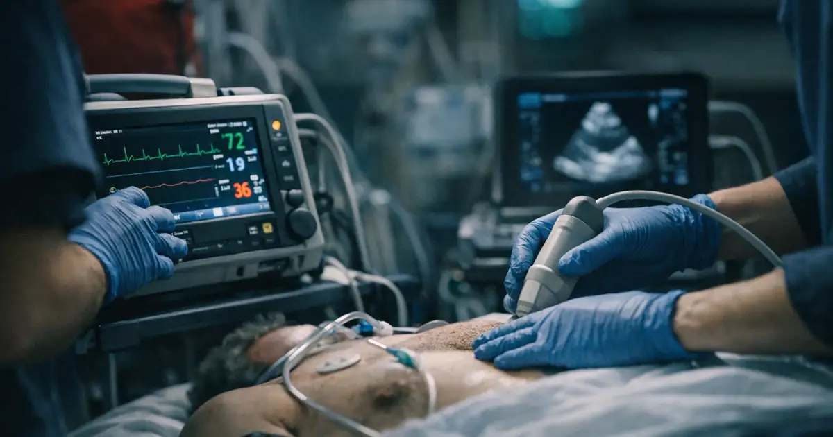
For the unstable patient, bedside ultrasound can answer time-critical questions while the team prepares imaging or treatment. It can support the working diagnosis and show acute right-heart pressure, but it does not replace the whole clinical assessment (Rouleau et al.).
For everyone else, pause before ordering a test. Ask what the pretest probability is and what result would change management. A D-dimer is a rule-out test for a selected population. It is not a substitute for clinical assessment, and a positive result does not tell you that the patient has a dangerous clot. The 2026 guideline addresses clinical diagnosis, risk assessment, adjunctive cardiovascular testing, and acute management as connected decisions (AHA/ACC guideline).
Rule out PE without reflex CT
Use a validated pathway that matches the patient. In a patient judged to have very low clinical probability, the Pulmonary Embolism Rule-out Criteria (PERC) can identify patients who need no D-dimer or imaging when all criteria are negative. PERC is not a universal screen for dyspnea, tachycardia, or chest pain. If probability is not low enough for PERC, move to D-dimer or imaging (2026 AHA/ACC/ACCP/ACEP/CHEST guideline).
If D-dimer is appropriate, use the assay's units and your local protocol. For patients older than 50 years, age-adjusted thresholds commonly use age multiplied by 10 ng/mL in fibrinogen-equivalent units. Probability-adapted approaches such as YEARS and PEGeD can reduce imaging in selected low-risk patients, but they do not perform the same way in every population. A 2024 emergency-department comparison found that age adjustment increased specificity without a significant sensitivity decrease in the older group, while YEARS and PEGeD had higher specificity but lower sensitivity in parts of the cohort (comparison of PE prediction rules).
That finding should change how residents present the result. Do not say, "The YEARS cutoff is 1,000, so the patient is safe." Say, "The patient met zero or one YEARS items, the assay units are..., the clinical probability is..., and this is the pathway our department follows." The number only has meaning inside the rule that produced it.
When imaging is indicated, choose the study with renal function, contrast risk, pregnancy status, prior imaging, and local expertise in mind.
After confirmation, let risk drive disposition
Once PE is confirmed, stop talking about the scan as if it answered the whole question. Ask whether the patient is hemodynamically stable, whether there is right-heart stress, whether bleeding risk changes anticoagulation, and whether the patient can manage treatment and follow-up outside the hospital. The 2024 review of low-risk PE describes the simplified Pulmonary Embolism Severity Index (sPESI) and Hestia criteria as tools that combine medical risk with practical discharge conditions (low-risk PE management review).
Home treatment is not the same as a quick discharge. It requires an anticoagulation plan, medication access, return precautions, follow-up, and a patient who can understand the plan. An individual-patient meta-analysis of 2,694 carefully selected home-treated patients found 30-day mortality of 0.30% and 30-day adverse events of 1.2%; cancer, abnormal troponin, and abnormal natriuretic peptide were associated with higher risk (home treatment meta-analysis). Those numbers describe selected patients. They do not make a score a replacement for judgment.
Intermediate-risk PE deserves a separate conversation. Right-ventricular dilation plus clinical risk factors may prompt consultation with critical care, cardiology, pulmonary, interventional radiology, or a local PE response team. The 2025 PEERLESS trial enrolled 550 patients with intermediate-risk PE, right-ventricular dilation, and added clinical risk factors. Large-bore thrombectomy beat catheter-directed thrombolysis on a hierarchical endpoint driven by deterioration, bailout, and intensive-care use, but the trial found no difference in mortality, intracranial hemorrhage, or major bleeding (PEERLESS randomized trial). That is a consultation signal, not permission to send every intermediate-risk patient for a procedure.
The most useful disposition note states the risk category, evidence, bleeding assessment, treatment plan, and follow-up owner. It also says what would make the plan unsafe. A deliberate-practice case in the EM-Sim scenario catalog can help a learner rehearse the decision before a real patient makes the question urgent.
Dr. Chet's Take
I have watched PE care move from a binary question—clot or no clot—to a risk conversation that starts before the scan. That is progress. The patient with shock needs a different room, team, and clock than the patient with a small clot and a reliable plan for home anticoagulation. The scan matters. It does not get to make the whole decision for us.
That being said, I still see two errors. Some clinicians order a D-dimer before deciding whether the patient is low risk enough for it. Others see right-heart strain and jump straight to a procedure without asking what the patient is doing now. The honest answer is that our tools are aids, not verdicts. Write down the physiology, the pretest probability, and the reason the chosen path fits. That discipline survives a busy shift. It is also the kind of clinical judgment discussed across Global MedOps Command, where technology stays in its proper role: support the clinician, never replace the clinician.
If you are the attending, make the risk statement out loud before the patient moves. If you are in the bay, ask who owns anticoagulation, follow-up, and the return plan. If the patient arrived after a prehospital evaluation, close the loop with the EMS team; EMS-MedSim is built around practicing those handoffs and decisions before the next call. A PE diagnosis deserves a risk plan that a second clinician can read and act on.
Key Takeaways
- Stabilize the patient with shock or cardiopulmonary failure while activating senior help and a reperfusion pathway (high-risk PE review).
- Use PERC only after a low-risk clinical assessment; use D-dimer pathways only when the result can safely rule out PE.
- Treat age-adjusted, YEARS, and PEGeD thresholds as parts of named algorithms, not as free-floating numbers (2024 comparison study).
- After a positive scan, document severity, right-heart risk, bleeding risk, treatment, disposition, and follow-up.
- Home treatment is for selected low-risk patients with a real medication and follow-up plan (home-treatment meta-analysis).
FAQ
When can the PERC rule rule out pulmonary embolism in the ED?
Use PERC only when your initial clinical assessment places the patient in a very low-risk group and all PERC criteria are negative. If the patient is not low risk, use a different validated diagnostic pathway rather than using PERC to avoid a test.
What is the age-adjusted D-dimer cutoff for pulmonary embolism?
For patients older than 50 years, many protocols use age multiplied by 10 ng/mL in fibrinogen-equivalent units. Confirm the assay units and follow the local protocol before applying the number (comparison of PE prediction rules).
Can a patient with pulmonary embolism go home from the ED?
Some hemodynamically stable patients can receive outpatient treatment after a validated low-risk assessment. The decision also requires medication access, bleeding review, reliable follow-up, and clear return instructions (low-risk PE management review).
What should the ED do for high-risk pulmonary embolism?
Treat high-risk PE as a resuscitation problem. Support oxygenation and circulation, obtain rapid senior and specialty input, and pursue primary reperfusion when indicated by the patient's instability and local protocol (high-risk PE review).
How should emergency clinicians use the 2026 PE guideline?
Use it as the current adult framework for diagnosis, severity classification, treatment, and follow-up. Then adapt the pathway to the patient's physiology, contraindications, resources, and local PE response process (2026 AHA/ACC/ACCP/ACEP/CHEST guideline).
If you're a emergency physician, EM resident, medical student, or APP trying to understand how AI will actually impact your clinical practice — not just the hype — I put together a free practical guide. You can download it here: AI in EM Survival Guide.
Sources
- Writing Committee Members et al. 2026 AHA/ACC/ACCP/ACEP/CHEST/SCAI/SHM/SIR/SVM/SVN Guideline for the Evaluation and Management of Acute Pulmonary Embolism in Adults
- Silva et al. Comparison of the accuracy of four diagnostic prediction rules for pulmonary embolism in patients admitted to the emergency department
- Luijten et al. Safety of treating acute pulmonary embolism at home: an individual patient data meta-analysis
- Han et al. JTH in Clinic: management of low-risk pulmonary embolism
- Jaber et al. Large-Bore Mechanical Thrombectomy Versus Catheter-Directed Thrombolysis in the Management of Intermediate-Risk Pulmonary Embolism: Primary Results of the PEERLESS Randomized Controlled Trial
- Rouleau et al. Management of high-risk pulmonary embolism in the emergency department: A narrative review
- Related simulation training: EMS-MedSim
- Books by Dr. Shermer