FAST Exam Mastery: A Practical Guide for Emergency Physicians
Chester "Chet" Shermer, MD, FACEP · May 19, 2026
About the author: Dr. Chester Shermer, MD, FACEP
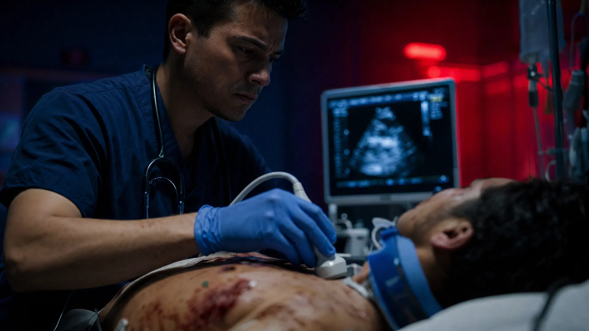
Master the FAST exam with this step-by-step guide for emergency physicians. Learn the four views, common pitfalls, and how POCUS changes resuscitation decisions in trauma.
The Focused Assessment with Sonography for Trauma (FAST) exam is a cornerstone of resuscitation in the emergency department. It's rapid, non-invasive, repeatable, and provides critical information that can significantly alter patient management. Mastery of the FAST exam is not just about identifying free fluid; it's about integrating those findings into a comprehensive clinical picture, understanding its limitations, and knowing when to escalate care based on its results.
The Four Standard FAST Views: What You're Looking For
The standard FAST exam systematically evaluates four anatomical regions for the presence of free intraperitoneal or pericardial fluid.
1. Pericardial View (Subxiphoid or Parasternal Long)
This is often the first view obtained, as pericardial tamponade is immediately life-threatening and readily reversible.
- Probe Position:
- Subxiphoid: Place the curvilinear or phased array probe just inferior to the xiphoid process, with the indicator pointing towards the patient's left shoulder. Angle the probe steeply towards the heart. This view is often challenging due to body habitus or patient discomfort.
- Parasternal Long Axis (PSLA): Place the phased array probe in the 3rd or 4th intercostal space, just to the left of the sternum, with the indicator pointing towards the patient's right shoulder. This view is often easier to obtain and provides excellent visualization of the pericardium.
- What You're Looking For:
- Pericardial Effusion: Anechoic (black) stripe between the visceral and parietal pericardium. Even a small amount can be significant in trauma due to the acute nature of fluid accumulation.
- Right Ventricular Collapse: Diastolic collapse of the right ventricle is a hallmark of tamponade.
- Left Ventricular Size/Function: Gross assessment of cardiac contractility can provide clues to hypovolemia or pre-existing cardiac disease.
- Clinical Pearl: In a hypotensive trauma patient, a small pericardial effusion with signs of right ventricular collapse demands immediate intervention (pericardiocentesis or surgical exploration). Do not wait for hemodynamic collapse to be profound.
2. Right Upper Quadrant (RUQ) View (Morison's Pouch)
This view assesses the interface between the liver and the right kidney (Morison's pouch), the right paracolic gutter, and the superior aspect of the right hemidiaphragm.
- Probe Position: Place the curvilinear probe in the mid-axillary line, between the 8th and 11th intercostal spaces, with the indicator pointing cephalad. Fan through the liver and kidney.
- What You're Looking For:
- Free Fluid in Morison's Pouch: Anechoic stripe between the liver and the right kidney. This is the most common location for free fluid to accumulate in supine patients due to gravity.
- Right Pleural Effusion: Anechoic fluid superior to the diaphragm, often with a "spine sign" (vertebrae visible above the diaphragm).
- Subcapsular Hematoma: Irregularity or anechoic/hypoechoic collection immediately beneath the liver capsule.
- Clinical Pearl: Morison's pouch is highly sensitive for free fluid. Even a thin anechoic stripe here in a hypotensive trauma patient is highly concerning for active hemorrhage.
3. Left Upper Quadrant (LUQ) View (Splenorenal Recess)
This view evaluates the interface between the spleen and the left kidney, the left paracolic gutter, and the superior aspect of the left hemidiaphragm.
- Probe Position: Place the curvilinear probe in the posterior axillary line, between the 6th and 9th intercostal spaces, with the indicator pointing cephalad. Fan through the spleen and kidney.
- What You're Looking For:
- Free Fluid in Splenorenal Recess: Anechoic stripe between the spleen and the left kidney.
- Left Pleural Effusion: Anechoic fluid superior to the diaphragm, with a "spine sign."
- Subcapsular Hematoma: Irregularity or anechoic/hypoechoic collection immediately beneath the splenic capsule.
- Clinical Pearl: The splenorenal recess is often a more challenging view due to the higher position of the spleen and overlying ribs. Patient inspiration can help drop the spleen into view. Remember that the spleen is more frequently injured than the liver in blunt abdominal trauma.
4. Pelvic View (Suprapubic)
This view assesses the rectovesical pouch in males and the rectouterine (Pouch of Douglas) and vesicouterine pouches in females.
- Probe Position: Place the curvilinear probe transversely just superior to the pubic symphysis, with the indicator pointing towards the patient's right. Then rotate 90 degrees for a sagittal view, with the indicator pointing cephalad.
- What You're Looking For:
- Free Fluid: Anechoic collection posterior to the bladder (Pouch of Douglas in females, rectovesical pouch in males) or anterior to the uterus (vesicouterine pouch in females).
- Bladder Integrity: Gross assessment of bladder wall for rupture, though this is not highly sensitive.
- Clinical Pearl: A full bladder acts as an acoustic window, making this view easier. If the bladder is decompressed, consider filling it with saline via a Foley catheter to improve visualization. Free fluid in the pelvis can be the only sign of significant intra-abdominal hemorrhage, especially in pelvic fractures.
The Extended FAST (eFAST) and When to Use It
The eFAST expands upon the standard FAST by adding evaluation of the bilateral pleural spaces for pneumothorax. This is crucial in trauma, especially in patients with chest wall injury or respiratory distress.
- Probe Position:
- Anterior Thoracic Views: Place the high-frequency linear probe in the 2nd-4th intercostal spaces along the midclavicular line bilaterally.
- Lateral Thoracic Views: Place the high-frequency linear probe in the 5th-7th intercostal spaces along the mid-axillary line bilaterally.
- What You're Looking For:
- Lung Sliding: The shimmering, to-and-fro movement of the visceral pleura against the parietal pleura with respiration. This rules out pneumothorax at the scanning site.
- B-lines: Vertical artifacts extending from the pleural line, often seen in pulmonary contusion or pulmonary edema.
- Absence of Lung Sliding: The primary sonographic sign of pneumothorax.
- "Lung Point": The specific location where lung sliding transitions to absent lung sliding, indicating the edge of the pneumothorax. This is highly specific for pneumothorax.
- "Stratosphere Sign" (M-mode): In M-mode, normal lung sliding creates a "seashore sign" (sandy appearance below the pleural line). Absence of lung sliding results in a "stratosphere sign" (parallel lines throughout).
- When to Use It: The eFAST should be performed as part of the primary survey in any trauma patient with suspected chest injury, respiratory distress, or significant mechanism of injury. It is particularly useful in hypotensive trauma patients to rapidly identify tension pneumothorax or massive hemothorax.
- Clinical Pearl: Always use a high-frequency linear probe for pneumothorax assessment. The curvilinear probe lacks the resolution to adequately visualize the pleural line and lung sliding. Scan multiple intercostal spaces bilaterally, as a pneumothorax may be localized.
Common Technical Pitfalls and How to Avoid Them
Even experienced sonographers can encounter challenges. Awareness of these pitfalls is key to accurate interpretation.
- Obesity/Body Habitus: Increased adipose tissue attenuates the ultrasound beam, making penetration difficult.
- Avoidance: Use a lower frequency curvilinear probe, apply more pressure, try different patient positions (e.g., left lateral decubitus for RUQ), and optimize gain/depth settings.
- Bowel Gas: Gas reflects ultrasound waves, creating artifact (dirty shadowing) that obscures deeper structures.
- Avoidance: Apply graded compression to displace gas, try different probe angles, scan during breath holds, and understand that bowel gas can mimic free fluid if misinterpreted.
- Subcutaneous Emphysema: Air in the subcutaneous tissues can prevent adequate visualization of the pleural line for pneumothorax assessment.
- Avoidance: Acknowledge the limitation and rely on other clinical signs or alternative imaging.
- Clotted Blood: Clotted blood can appear hyperechoic or isoechoic, making it difficult to distinguish from solid organs or normal tissue.
- Avoidance: Look for characteristic shapes of fluid collections, track fluid into dependent areas, and correlate with clinical findings.
- Pre-existing Fluid: Ascites, peritoneal dialysis fluid, or pre-existing pericardial effusions can be mistaken for traumatic hemorrhage.
- Avoidance: Obtain a thorough history if possible. In the acute trauma setting, assume new fluid is traumatic until proven otherwise, especially if the patient is unstable.
- Mirror Artifact: Can occur in the RUQ, creating a "phantom" liver above the diaphragm, which can obscure a true pleural effusion.
- Avoidance: Recognize the characteristic appearance; the artifactual image is a mirror of the real image. Look for the "spine sign" to confirm pleural fluid.
- Misinterpreting Perinephric Fat: The hyperechoic fat surrounding the kidney can be mistaken for free fluid, especially in the splenorenal view.
- Avoidance: Free fluid is anechoic (black) and conforms to spaces. Perinephric fat is hyperechoic and has a distinct texture.
- Insufficient Fanning: Not adequately fanning through the entire organ or recess can lead to missing small fluid collections.
- Avoidance: Systematically fan through each view, ensuring complete visualization.
How FAST Findings Change Your Resuscitation Algorithm in Penetrating vs. Blunt Trauma
The interpretation and subsequent management based on FAST findings differ significantly between blunt and penetrating trauma.
Blunt Trauma
- Positive FAST (Free Fluid): In a hemodynamically unstable blunt trauma patient, a positive FAST indicates intra-abdominal hemorrhage requiring immediate surgical intervention (laparotomy). This is a "go to the OR" decision.
- Negative FAST: In a hemodynamically unstable blunt trauma patient, a negative FAST (especially if repeated) should prompt a search for other sources of hemorrhage (e.g., retroperitoneal, pelvic, long bone fractures, massive hemothorax) or non-hemorrhagic causes of shock (e.g., neurogenic, cardiogenic, tension pneumothorax). If no other source is found, consider a repeat FAST, CT scan (if stable enough), or diagnostic peritoneal lavage (DPL) if CT is unavailable or contraindicated.
- Positive FAST (Free Fluid) in a Hemodynamically Stable Patient: This typically warrants further imaging, usually a CT scan of the abdomen and pelvis with IV contrast, to characterize the injury, quantify the fluid, and identify specific organ damage. Surgical intervention may still be necessary, but it is not as immediate as in the unstable patient.
- eFAST for Pneumothorax/Hemothorax: A positive eFAST for pneumothorax (especially tension) or massive hemothorax in an unstable patient mandates immediate tube thoracostomy.
Penetrating Trauma
- Positive FAST (Pericardial Fluid): In any penetrating chest trauma, even a small pericardial effusion, especially with signs of tamponade, is an indication for immediate surgical exploration (thoracotomy/sternotomy) or pericardiocentesis as a temporizing measure. This is a "go to the OR" decision.
- Positive FAST (Intraperitoneal Fluid): In penetrating abdominal trauma, a positive FAST often indicates significant intra-abdominal injury and is usually an indication for immediate surgical exploration (laparotomy), especially if the patient is unstable. However, even in stable patients, a positive FAST in penetrating trauma is often treated more aggressively than in blunt trauma due to the higher likelihood of hollow viscus injury and peritonitis.
- Negative FAST in Penetrating Trauma: A negative FAST in penetrating trauma, particularly stab wounds, is less reassuring than in blunt trauma. It does not rule out significant injury, especially hollow viscus perforation, diaphragm injury, or retroperitoneal injury. Further evaluation (local wound exploration, serial abdominal exams, CT scan, DPL, diagnostic laparoscopy) is often necessary, even in stable patients.
- eFAST for Pneumothorax/Hemothorax: Similar to blunt trauma, a positive eFAST for pneumothorax or hemothorax in penetrating chest trauma requires immediate intervention (chest tube).
Sensitivity/Specificity Limitations Every EP Must Know
While invaluable, the FAST exam has inherent limitations that must be understood to avoid diagnostic errors.
- Sensitivity for Free Fluid:
- The FAST exam is highly operator-dependent.
- It typically requires at least 200-600 mL of free fluid to be reliably detected. Smaller amounts, especially in the early stages of hemorrhage, can be missed.
- Sensitivity is lower for isolated retroperitoneal hemorrhage (e.g., renal injury, aortic injury) because these spaces are not directly assessed by the standard FAST.
- Sensitivity for specific organ injury (e.g., splenic laceration, liver laceration) is low. It detects the result of the injury (free fluid), not the injury itself.
- Specificity for Free Fluid:
- Specificity is generally high for free fluid in the trauma setting. However, pre-existing ascites or peritoneal dialysis fluid can lead to false positives.
- The FAST cannot distinguish between blood, urine, bile, or bowel contents. In the trauma setting, free fluid is assumed to be blood until proven otherwise.
- Limitations in Detecting Specific Injuries:
- Hollow Viscus Injury: The FAST exam is very poor at detecting hollow viscus injury (e.g., bowel perforation) because the amount of free fluid (bowel contents, air) may be minimal and not easily visualized.
- Diaphragmatic Injury: Difficult to detect directly.
- Pancreatic/Duodenal Injury: Often retroperitoneal and may not produce free intraperitoneal fluid.
- Mesenteric Injury: Can cause significant hemorrhage without free fluid or with only localized hematoma.
- Limitations for Pneumothorax:
- The eFAST is highly sensitive for anterior pneumothoraces.
- It can miss small or apical pneumothoraces, especially if not adequately scanned.
- Subcutaneous emphysema can obscure the pleural line, rendering the exam uninterpretable for pneumothorax.
When a Negative FAST Is Not Reassuring
A negative FAST exam does not always mean the patient is free of significant injury. Several scenarios warrant continued vigilance and further investigation:
- Hemodynamically Unstable Patient with Negative FAST: This is a critical situation. A negative FAST in an unstable patient means the source of shock is not readily apparent from the abdominal or pericardial views. This should prompt:
- Repeat FAST: Small amounts of fluid may not be present initially but can accumulate.
- Search for other sources of hemorrhage: Pelvic fractures, retroperitoneal hemorrhage, long bone fractures, massive hemothorax (if eFAST not performed or negative).
- Search for non-hemorrhagic causes of shock: Tension pneumothorax (if eFAST negative), neurogenic shock, cardiogenic shock, anaphylaxis, sepsis.
- Consider DPL: In select cases where CT is unavailable or contraindicated, and the source of instability remains elusive.
- Penetrating Trauma with Negative FAST: As discussed, a negative FAST in penetrating trauma, especially stab wounds to the abdomen or flank, does not rule out significant hollow viscus injury, diaphragm injury, or retroperitoneal injury. These patients often require serial abdominal exams, local wound exploration, CT imaging, or diagnostic laparoscopy.
- High-Risk Mechanism of Injury with Negative FAST: Patients with severe mechanisms (e.g., high-speed MVC, fall from height, pedestrian struck) despite a negative FAST, still warrant a high index of suspicion for occult injury. If stable, these patients generally proceed to CT imaging.
- Altered Mental Status/Intoxication with Negative FAST: Patients who cannot reliably report symptoms or have a compromised neurological exam require a lower threshold for further imaging, even with a negative FAST, as they may mask signs of peritonitis or pain.
- Delayed Presentation: Patients presenting hours after trauma may have had time for small bleeds to clot or for fluid to accumulate to detectable levels. A negative FAST in the immediate post-trauma period may become positive later. Serial exams are crucial.
- Suspected Retroperitoneal Injury: The FAST exam does not effectively evaluate the retroperitoneal space. Renal trauma, major vascular injury (e.g., aortic dissection), or pancreatic injury can cause significant hemorrhage or inflammation without free intraperitoneal fluid.
- Suspected Diaphragmatic Injury: This is a difficult diagnosis with FAST.
Mastery of the FAST exam is an ongoing process. It requires not only technical proficiency but also a deep understanding of its physiological basis, clinical context, and inherent limitations. Integrate it thoughtfully into your trauma resuscitation, but never let a negative FAST overshadow a strong clinical suspicion for injury.
Practice Your FAST Exam Skills in Simulation
Reading about the FAST exam is one thing — executing it correctly under pressure is another. EM-Sim's Penetrating Chest Trauma with Tension Pneumothorax scenario puts you in a high-stakes trauma bay where POCUS findings directly change your management algorithm.
Try the Free Scenario — No Login Required →
Or explore the full scenario catalog for more ultrasound-integrated cases: