Managing the Undifferentiated Hypotensive Patient: A Systematic Approach
Chester "Chet" Shermer, MD, FACEP · May 18, 2026
About the author: Dr. Chester Shermer, MD, FACEP
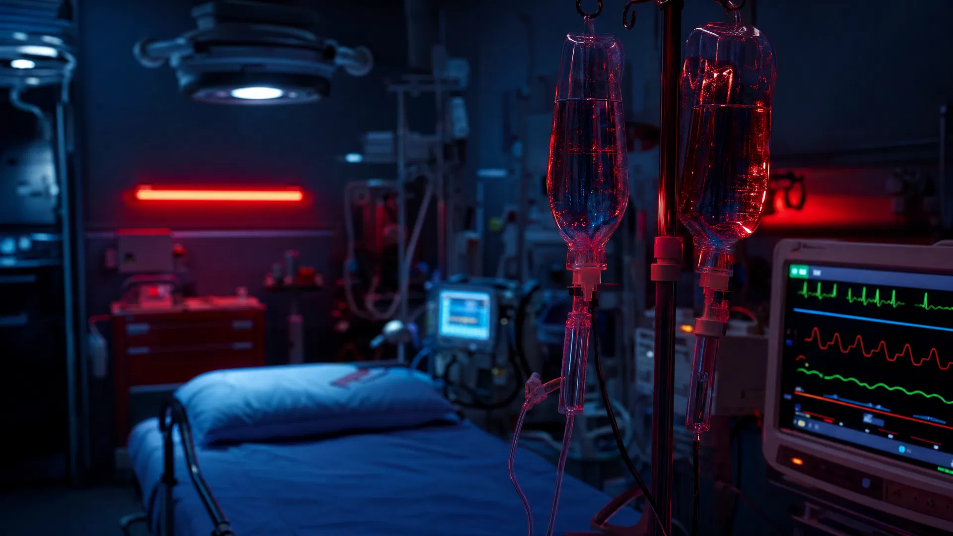
When a hypotensive patient rolls through your ED doors, your first 10 minutes determine outcomes. Here's a systematic framework for the undifferentiated hypotensive patient.
When a hypotensive patient rolls through your ED doors, your first 10 minutes determine outcomes. The stakes are incredibly high, the differential vast, and the need for rapid, accurate diagnosis and intervention paramount. The undifferentiated hypotensive patient is one of the most challenging and rewarding presentations we face in emergency medicine. This isn't just about giving fluids or starting a pressor; it's about a systematic, simultaneous approach to resuscitation, diagnosis, and ongoing management that can literally mean the difference between life and death.
The Immediate Assessment: ABCs and the "Shock Clock"
Before you even consider a differential, focus on the immediate threats. This is the "Shock Clock" – the critical first few minutes where you stabilize, assess, and initiate life-saving interventions.
- Airway: Is it patent? Is the patient protecting it? Are they speaking in full sentences? If not, consider airway adjuncts or definitive airway management.
- Breathing: Is the patient tachypneic? Bradypneic? Are breath sounds equal? Is there accessory muscle use? Obtain an SpO2 and consider capnography.
- Circulation: This is where hypotension takes center stage.
- Rapid Vitals: Get a blood pressure, heart rate, respiratory rate, temperature, and SpO2.
- Access: Establish at least two large-bore IVs (18G or larger, ideally 16G). If peripheral access is difficult, consider intraosseous (IO) access immediately. Don't waste precious minutes trying to "find a vein."
- Labs: Draw a full set of labs: CBC, chemistry panel (electrolytes, creatinine, BUN, glucose), lactate, ABG/VBG, coagulation studies, cardiac enzymes, type and screen, and cultures (blood, urine, sputum if indicated).
- ECG: Get a 12-lead ECG immediately to rule out acute coronary syndromes or significant arrhythmias.
- Fluid Bolus: Unless there's clear evidence of cardiogenic shock or fluid overload (e.g., acute pulmonary edema), administer a rapid 500 mL to 1 Liter bolus of crystalloid (e.g., Lactated Ringer's or Plasma-Lyte). Reassess after each bolus. Remember, fluids are a drug; they have indications, contraindications, and side effects.
- Point-of-Care Ultrasound (POCUS): This is your most powerful diagnostic tool in the initial assessment. More on this below.
The POCUS-Driven Differential: RUSH Exam
The RUSH (Rapid Ultrasound for Shock and Hypotension) exam is a game-changer. It allows you to quickly categorize shock at the bedside, often within minutes, guiding your resuscitation strategy. I teach this extensively at Global MedOps Command because it directly impacts patient outcomes.
The RUSH exam systematically assesses:
- Pump (Cardiac):
- Pericardial Effusion/Tamponade: Look for fluid around the heart and diastolic collapse of the right ventricle.
- LV Function: Gross assessment of contractility (hyperdynamic, normal, hypodynamic, or severely depressed).
- RV Strain: Look for RV dilation and dysfunction, often seen in massive pulmonary embolism.
- Tank (Fluid Status/Volume):
- IVC Collapsibility: A small, collapsing IVC suggests hypovolemia. A plethoric, non-collapsing IVC suggests fluid overload or obstructive shock. Caveat: IVC assessment can be unreliable in intubated, ventilated patients or those with elevated intra-abdominal pressure.
- Free Fluid: Look for free fluid in the abdomen (FAST exam windows) or pleural effusions.
- Pipes (Vascular Tone/Obstruction):
- Aorta: Look for abdominal aortic aneurysm (AAA) or dissection.
- Lungs (part of the "Tank" assessment): Look for B-lines (suggesting tank overload / pulmonary edema) or pneumothorax (absence of lung sliding, barcode sign on M-mode).
- DVT: While not part of the standard RUSH, if PE is suspected, quickly scan for proximal DVT.
Based on your RUSH findings, you can often narrow down your differential into the major categories of shock:
- Hypovolemic Shock: Small, collapsing IVC; hyperdynamic LV; no pericardial effusion; no RV strain; no significant free fluid (unless traumatic hemorrhage).
- Cardiogenic Shock: Poor LV function; dilated, non-collapsing IVC; B-lines on lung ultrasound.
- Obstructive Shock:
- Tamponade: Pericardial effusion with RV diastolic collapse.
- Massive PE: RV dilation/dysfunction; small, collapsing LV; dilated IVC; +/- DVT.
- Tension Pneumothorax: Absence of lung sliding, barcode sign on M-mode; tracheal deviation (clinical).
- Distributive Shock: Hyperdynamic LV; dilated, non-collapsing IVC (often initially, then variable); no pericardial effusion; no RV strain; no free fluid.
The Differential Diagnosis and Targeted Interventions
Once you've categorized the shock, your interventions become more targeted.
Hypovolemic Shock
- Causes: Hemorrhage (GI bleed, trauma, ruptured AAA, ectopic pregnancy), severe dehydration (vomiting, diarrhea, burns, DKA).
- Management:
- Fluids: Aggressive crystalloid resuscitation (e.g., 1-2 Liters rapidly, then reassess).
- Blood Products: If hemorrhagic, activate massive transfusion protocol (MTP) early. Target a 1:1:1 ratio of PRBCs, FFP, and platelets. Consider tranexamic acid (TXA) for trauma or significant hemorrhage within 3 hours of injury.
- Source Control: Identify and stop the bleeding (e.g., surgery for trauma, endoscopy for GI bleed, IR embolization).
Cardiogenic Shock
- Causes: Acute MI, severe heart failure exacerbation, valvular emergencies, myocarditis, arrhythmias.
- Management:
- Avoid Aggressive Fluids: Be judicious with fluids; often small boluses (250 mL) or even diuretics are needed.
- Inotropes/Vasopressors: Norepinephrine is the preferred first-line vasopressor; dopamine is associated with more arrhythmias and worse outcomes in cardiogenic shock (SOAP II). Consider dobutamine or milrinone for severe LV dysfunction.
- Treat Underlying Cause: Reperfusion for STEMI, antiarrhythmics for arrhythmias, afterload reduction if appropriate.
- Consult Cardiology: Early consultation for potential mechanical circulatory support (IABP, Impella, ECMO).
Obstructive Shock
- Causes: Cardiac tamponade, massive pulmonary embolism, tension pneumothorax.
- Management:
- Cardiac Tamponade: Emergent pericardiocentesis (ultrasound-guided).
- Massive PE: Thrombolysis (systemic or catheter-directed), embolectomy, vasopressors (norepinephrine) to maintain RV perfusion.
- Tension Pneumothorax: Needle decompression followed by chest tube insertion.
Distributive Shock
- Causes: Sepsis, anaphylaxis, neurogenic shock, adrenal crisis.
- Management:
- Sepsis:
- Fluids: Initial fluid bolus (30 mL/kg crystalloid) within the first 3 hours, then reassess. Avoid excessive fluids if not improving.
- Vasopressors: Norepinephrine is first-line. Add vasopressin or epinephrine if needed.
- Antibiotics: Broad-spectrum antibiotics within 1 hour of recognition of sepsis.
- Source Control: Identify and treat the infection.
- Anaphylaxis: Epinephrine (IM first, then IV infusion if severe), H1/H2 blockers, steroids, fluids.
- Neurogenic Shock: Vasopressors (norepinephrine, phenylephrine) to maintain mean arterial pressure (MAP) and spinal cord perfusion. Avoid excessive fluids.
- Adrenal Crisis: Stress-dose hydrocortisone, fluids.
Ongoing Monitoring and Reassessment
Resuscitation is not a one-time event; it's a continuous cycle of intervention and reassessment.
- Frequent Vitals: Every 5-15 minutes, depending on stability.
- Urine Output: Insert a Foley catheter to monitor urine output (target >0.5 mL/kg/hr).
- Lactate Clearance: Trend lactate levels. A decreasing lactate indicates effective resuscitation.
- Mental Status: Improving mental status is a good sign of cerebral perfusion.
- Repeat POCUS: Re-evaluate cardiac function, IVC, and lung fields after interventions.
- Arterial Line: Consider an arterial line for continuous, accurate blood pressure monitoring and frequent blood gas sampling in critically ill patients.
- Central Venous Catheter: While not universally required, CVCs can be useful for vasopressor administration and CVP monitoring (though CVP as a sole guide to fluid responsiveness is limited).
The Role of Simulation in Mastering Shock Management
Managing the undifferentiated hypotensive patient is a high-acuity, low-frequency event for many clinicians, yet it demands immediate, expert action. This is precisely where simulation-based training excels. At Global MedOps Command, our EM-Sim programs immerse participants in realistic scenarios where they must:
- Rapidly assess and stabilize: Practice the "Shock Clock" under pressure.
- Perform POCUS: Develop proficiency in the RUSH exam to guide diagnosis.
- Make critical decisions: Choose appropriate fluids, vasopressors, and other interventions.
- Manage a team: Practice communication, delegation, and leadership in a crisis.
- Handle complications: Address decompensation, medication errors, or procedural difficulties in a safe environment.
Simulation allows you to make mistakes, learn from them, and refine your approach without patient harm. It builds the muscle memory and cognitive frameworks necessary to perform flawlessly when it matters most. Mastering the systematic approach to shock management through deliberate practice in simulation translates directly to improved patient care and confidence in the emergency department.
Chet Shermer, MD, FACEP, is the Founder & Medical Director of Global MedOps Command. He is a board-certified emergency physician with over two decades of clinical experience and a passion for advancing medical education through simulation. Learn more about EM-Sim programs at emsim.globalmedopscommand.com.
Practice the Undifferentiated Hypotensive Patient on EM-Sim
The systematic approach described above is only as effective as your ability to execute it under pressure. EM-Sim's Multi-System Trauma with Hemorrhagic Shock and Septic Shock Recognition scenarios force you to work through the exact differential described in this article — with branching decision trees that mirror real resuscitation complexity.